ÜMMÜYE BİÇER: NECMETTİN ERBAKAN ÜNİ. MERAM TIP FAK.
DURDU MEHMET YAVŞAN: NECMETTİN ERBAKAN ÜNİVERSİTESİ
SONER DEMİRBAŞ: SELÇUK ÜNİVERSİTESİ
TURGUT TEKE: NECMETTİN ERBAKAN ÜNİVERSİTESİ
Year : 2016
Vol : 32
Issue : Ek
Page : 8-11
Eosinophilic pulmonary diseases (EPD) are defined as a group of
diseases characterized by the in crease of eosinophils in the air way
and/or lung tissue. While a part of the sedis orders mainly affects the
pulmonary, others show systemic in volvement. Chronical eosinophilic
pneumonia (CEP) should be kept in mind in the existence of subacute
or chronic respiratory and general symptoms, alveoler and/or bloode
osinophilia and peripheral pulmonary in filtrates on chest radiography.
A 53-year-old male patient was admitted to our clinic with on going
complaints of cough, occasional fever and malaise for six months.
When two-sided apical, subapical and the infiltration around hiluses
were detected on chest x-ray, the case was hospitalized with the
prediagnosis of pulmonary tuberculosis. Eosinophilia in hemogram,
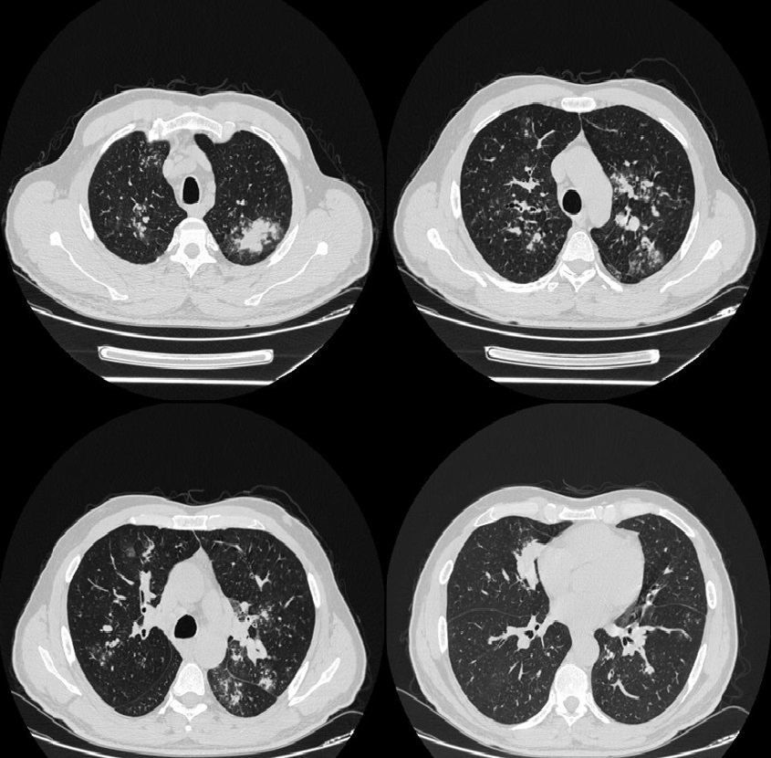
bilateral infiltrative lesions with irregular contours and bilateral cystic
bronc hiectasis foci were detected on high resolution computed
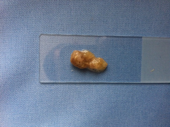
tomography (HRCT). Bronchoscopy revealed purulent secretions and
thick mucous in hibations. No specific etiologic cause was defined
in the etiology, and the case wase valuated as CEP. Due to unusual
bronchoscopic and radiological findings, the case was presented and
discussed.
 Case Report
 View as PDF
 Keywords
Chronic eosinophilic pneumonia, high resolution computed tomography, bronchoscopy
Keywords
Chronic eosinophilic pneumonia, high resolution computed tomography, bronchoscopy