Diagnostic Utility of the Corpus Callosum Index in Mild and Moderate Alzheimer’s Disease and Its Relationship with Mini Mental State Examination Subcomponents
ORUÇ ŞAHİN
,Emine Hande Kılıçaslan Şahin
ORUÇ ŞAHİN: AKSARAY ÜNİVERSİTESİ TIP FAKÜLTESİ
Emine Hande Kılıçaslan Şahin: Beyhekim Eğitim ve Araştırma Hastanesi
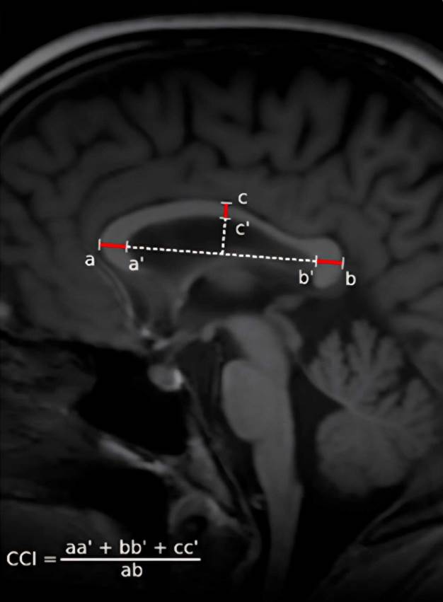
Objective: This study evaluated the utility of the corpus callosum index (CCI) as a discriminative biomarker between the mild and moderate stages of Alzheimer’s
disease (AD).
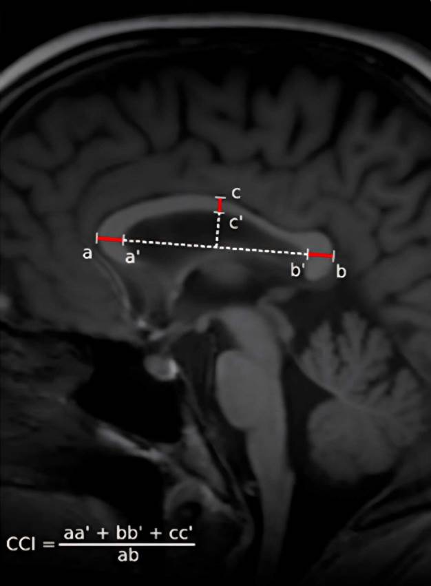
Materials and Methods: A total of 168 individuals were included, comprising two patient groups diagnosed with mild and moderate AD and a control group. CCI
values and Mini-Mental State Examination (MMSE) subcomponent scores were compared. Statistical analyses assessed correlations between MMSE subdomains and
CCI, group differences, optimal cutoff values using receiver operating characteristic (ROC) analysis, and logistic regression models.
Results: Significant differences in CCI values were observed among the groups (p < 0.001). CCI values decreased progressively with increasing dementia severity. A
positive correlation was identified between CCI and total MMSE score, as well as orientation, attention-calculation, and language subtests. In the univariate logistic
regression analysis, age, CCI, and education level were significant variables; in the multivariate logistic regression model, both CCI and age remained independent
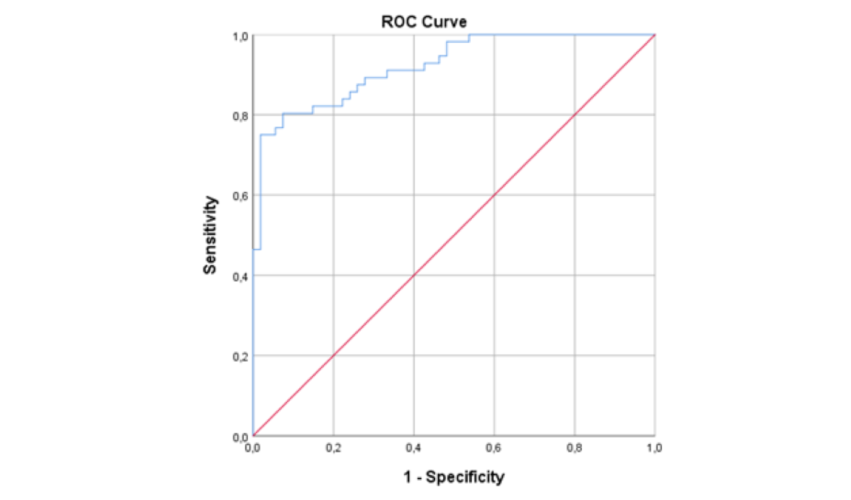
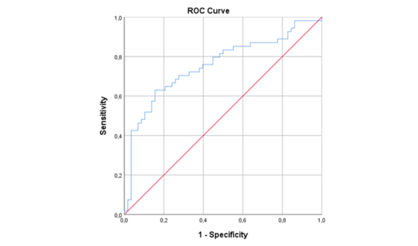
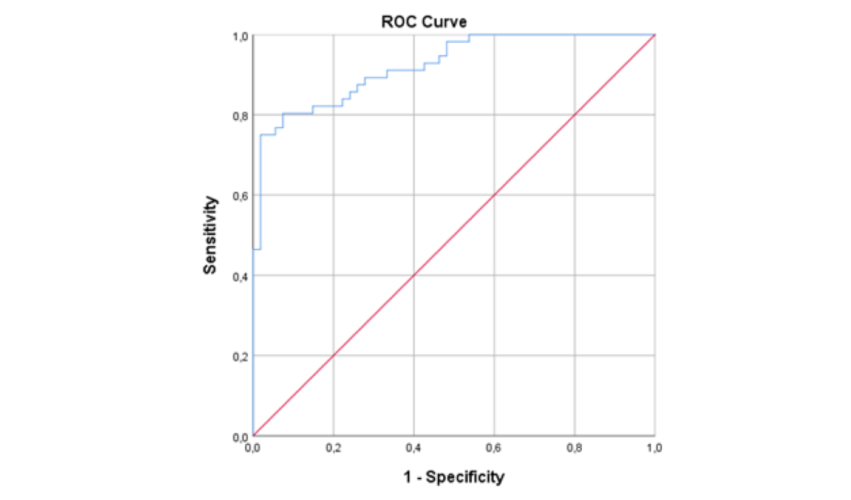
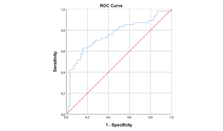
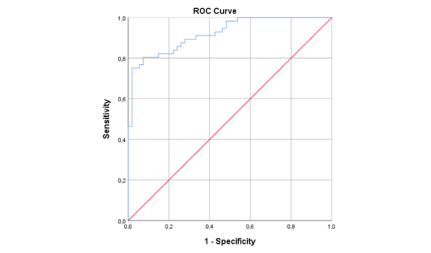
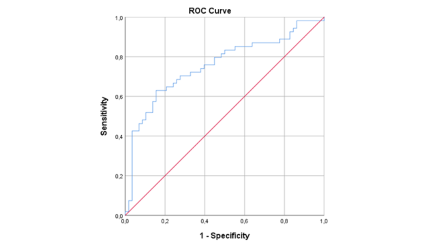
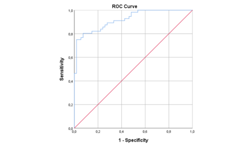
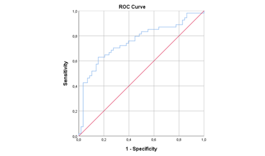
predictors of dementia severity (p < 0.001 for both). In the ROC analysis, CCI demonstrated high discriminative power between controls and patients with mild
dementia (AUC = 0.922) and acceptable discriminative power between mild and moderate dementia (AUC = 0.756).
Conclusion: The findings suggest that CCI may be a useful measure for the early assessment of AD severity. Its high sensitivity in distinguishing controls from patients
with mild dementia indicates potential clinical utility as an adjunctive assessment tool. However, larger multicenter prospective studies are needed to draw definitive
conclusions regarding the prognostic value and clinical applicability of CCI.
Cite this Article As :
Sahin O, Kilicaslan Sahin EH. Diagnostic Utility of the Corpus Callosum Index in Mild and Moderate Alzheimer’s Disease and Its Relationship with Mini
Mental State Examination Subcomponents. Selcuk Med J 2026;42(3): 252-260
1. Buathong S, Piyapitayanan S, Thientunyakit T, et al. Quantitative susceptibility mapping for Alzheimer’s disease, mild cognitive impairment, and normal aging: evaluation of corpus callosum. Quant Imaging Med Surg. 2025;15(5):4566-79. doi:10.21037/qims-24-319.
2. Ballard C, Gauthier S, Corbett A, et al. Alzheimer’s disease. Lancet. 2011;377:1019‑31. doi:10.1016/S0140-6736(10)61349-9.
3. Aillaud I, Funke SA, Tau aggregation inhibiting peptides as potential therapeutics for Alzheimer disease. Cell Mol Neurobiol. 2023;43:951‑61. doi:10.1007/s10571-022-01230-7.
4. Van Schependom J, Niemantsverdriet E, Smeets D, et al. Callosal circularity as an early marker for Alzheimer’s disease. Neuroimage Clin. 2018;19:516‑26. doi:10.1016/j.nicl.2018.05.018.
5. Ardekani BA, Bachman AH, Figarsky K, et al. Corpus callosum shape changes in early Alzheimer’s disease: an MRI study using the OASIS brain database. Brain Struct Funct. 2014;219(1):343‑52. doi:10.1007/s00429-013-0503-0.
6. Das S, Panigrahi P, Chakrabarti S, et al. Corpus callosum atrophy in detection of mild and moderate Alzheimer’s disease using brain magnetic resonance image processing and machine learning techniques. J Alzheimers Dis Rep. 2021;5(1):771‑88. doi:10.3233/ADR-210314.
7. Di Paola M, Spalletta G, Caltagirone C, et al. In vivo structural neuroanatomy of corpus callosum in Alzheimer’s disease and mild cognitive impairment using different MRI techniques: a review. J Alzheimers Dis. 2010;20(1):67‑95. doi:10.3233/JAD-2010-1370.
8. Zhou X, Li H, Li T, et al. Altered callosal morphology in post-stroke cognitive impairment. Brain Res. 2025;1847:149307. doi:10.1016/j.brainres.2024.149307.
9. Liu Y, Chen S, Li S, et al. Diagnostic and inclusion criteria in Alzheimer’s disease clinical trials: a systematic review of the past decade. J Alzheimers Dis Rep. 2025;9:25424823251362444. doi:10.1177/25424823251362444.
10. McKhann G, Drachman D, Folstein M, et al. Clinical diagnosis of Alzheimer’s disease: report of the NINCDS‑ADRDA Work Group under the auspices of Department of Health and Human Services Task Force on Alzheimer’s Disease. Neurology. 1984;34(7):939‑44. doi:10.1212/WNL.34.7.939.
11. Reisberg B, Vadukapuram R, Kenowsky S. The global deterioration scale (GDS). World Alzheimer Report. 2022:44. https://www.alzint.org/resource/world-alzheimer-report-2022/
12. Mavioglu H, Gedizlioglu M, Akyel S, et al. The validity and reliability of the Turkish version of Alzheimer’s Disease Assessment Scale–Cognitive Subscale (ADAS‑Cog) in patients with mild and moderate Alzheimer’s disease and normal subjects. Int J Geriatr Psychiatry. 2006;21(3):259‑65. doi:10.1002/gps.1457.
13. Gonçalves LI, Dos Passos GR, Conzatti LP, et al. Correlation between the corpus callosum index and brain atrophy, lesion load, and cognitive dysfunction in multiple sclerosis. Mult Scler Relat Disord. 2018;20:154‑58. doi:10.1016/j.msard.2018.01.015.
14. Liu M, Zhang J, Zong L, et al. The differential diagnostic value of the callosal angle and Evans index in mild cognitive impairment and Alzheimer’s disease. Curr Med Imaging Rev. 2021;17(7):889‑96. doi:10.2174/1573405616666201223150004.
15. Raybaud C. The corpus callosum, the other great forebrain commissures, and the septum pellucidum: anatomy, development, and malformation. Neuroradiology. 2010;52(6):447‑77. doi:10.1007/s00234-010-0696-3.
16. Valenzuela‑Fuenzalida JJ, Orellana‑Hidalgo S, Baeza‑Garrido V, et al. A systematic review with a meta‑analysis of the morphological variants of the corpus callosum: related neurocognitive clinical implications. World Neurosurg. 2025;124:317. doi:10.1016/j.wneu.2025.124317.
17. Liu X, Xu D, Zhong X, et al. Altered callosal morphology and connectivity in asymptomatic carotid stenosis. J Magn Reson Imaging. 2024;59(3):998‑07. doi:10.1002/jmri.28872.
18. Wang XD, Ren M, Zhu MW, et al. Corpus callosum atrophy associated with the degree of cognitive decline in patients with Alzheimer’s dementia or mild cognitive impairment: a meta‑analysis of the region of interest structural imaging studies. J Psychiatr Res. 2015;63:10‑19. doi:10.1016/j.jpsychires.2015.02.005.
19. Dogra H, Ding S, Yeon M, et al. Confounder adjustment in shape‑on‑scalar regression model: corpus callosum shape alterations in Alzheimer’s disease. Stats. 2023;6(4):980‑89. doi:10.3390/stats6040061.
20. Shaji S, Palanisamy R, Swaminathan R, et al. Structural irregularities in MR corpus callosal images and their association with cerebrospinal fluid biomarkers in mild cognitive impairments. Neurosci Lett. 2023;810:137329. doi:10.1016/j.neulet.2023.137329.
21. Talwar P, Kushwaha S, Chaturvedi M, et al. Systematic review of different neuroimaging correlates in mild cognitive impairment and Alzheimer’s disease. Clin Neuroradiol. 2021;31(4):953‑67. doi:10.1007/s00062‑021‑01057‑7.



 Keywords