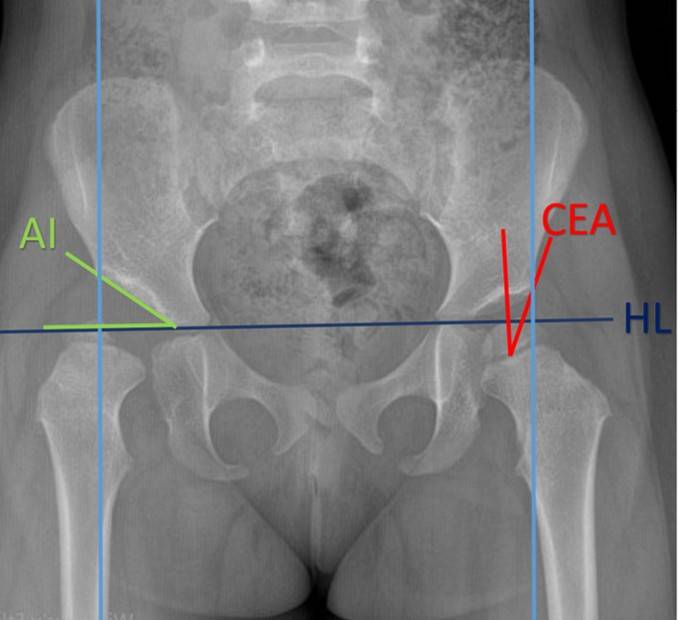
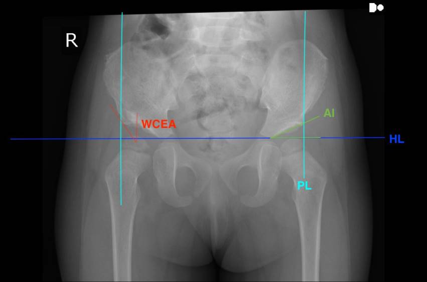
ABSTRACT Aim: Early diagnosis and timely care is important in patients with Developmental Hip Dysplasia (DDH). Late presenting or neglected patients may require various osteotomy procedures. There is a limited number of articles in the literature on Pembersal osteotomy in DDH surgery. The aim of this study was to assess the outcomes of isolated Pembersal osteotomy in children over 18 months of age with DDH. Materials and Methods: A retrospective analysis was made of the outcomes of patients who underwent Pembersal osteotomy for DDH at our clinic from 2014 to 2021. The study included 9 children (7 girls, 2 boys) aged between 19 and 36 months with at least 2 years of follow-up who underwent isolated periacetabular Pembersal osteotomy with open reduction. Preoperative, early postoperative, 6 weeks postoperative and final follow-up radiographs were evaluated and Wiberg's centre-edge angle (CEA), the acetabular index (AI) and Tönnis grades were noted. Results: The mean age of the patients was 28.75 months and the follow-up period was 50.33 months. CEA and AI showed improvement in the early postoperative period and this improvement continued in the later periods. Tönnis staging showed postoperative improvement in all patients, and this improvement continued in the early postoperative period and on the 6-week follow-up radiographs. At the final follow-up examination, 3 hips had CEA <20º, AI >20º and Tönnis stage ≥2 and these 3 hips were considered as a radiologically poor outcome. Conclusion: This study demonstrated that isolated Pembersal osteotomy is an effective and safe option for the treatment of developmental dysplasia of the hip, with improvement in acetabular index and other radiological parameters. Keywords: Developmental dysplasia of the hip, periacetabular osteotomy, Pembersal osteotomy, centre-edge angle, acetabular index
Cite this Article As :
Baymurat AC, Abdulaliyev F, Ahmadov A, Osmanli E, Ok N, Gul O, Turanli S. Early and Mid-Term Radiological Results of Isolated Pembersal Procedure in Cases of Developmental Dislocation of The Hip. Selcuk Med J 2024;40(1): 16-21
1. Bakarman K, Alsiddiky AM, Zamzam M, et al. Developmental Dysplasia of the Hip (DDH): Etiology, Diagnosis, and Management. Cureus. 2023; 15:43207.
2. Ponseti IV. Growth and development of the acetabulum in the normal child. Anatomical, histological, and roentgenographic studies. J Bone Joint Surg Am. 1978; 60:575-85
3. Sungur I, Cetinus ME. Treatment of developmental dysplasia of the hip in children older than 18 months. TOTBID Journal 2014; 13:412-425.
4. Şenar M, Westhoff B, Zilkens C, et al. Indikation und Ergebnisse hüftnaher Osteotomien bei Dysplasie [Indications and results of corrective pelvic osteotomies in developmental dysplasia of the hip]. Orthopade. 2008; 37:556-70, 572-4, 576. German.
5. Agarwal A, Rastogi P. Clinicoradiological outcomes following pembersal acetabular osteotomy for developmental dysplasia of hip in young children: A series of 16 cases followed minimum 2 years. J Clin Orthop Trauma. 2021; 23:101669.
6. Bursali A, Tonbul M. How are outcomes affected by combining the Pemberton and Salter osteotomies? Clin Orthop Relat Res. 2008; 466:837-46.
7. Perlik PC, Westin GW, Marafioti RL. A combination pelvic osteotomy for acetabular dysplasia in children. J Bone Joint Surg Am. 1985; 67:842-50.
8. Li Y, Guo Y, Li M, et al. Multi-center Pediatric Orthopedic Study Group of China. Acetabular index is the best predictor of late residual acetabular dysplasia after closed reduction in developmental dysplasia of the hip. Int Orthop. 2018; 42:631-640.
9. Weinstein SL, Mubarak SJ, Wenger DR. Developmental hip dysplasia and dislocation: Part I. Instr Course Lect. 2004; 53:523-30.
10. Wenger DR, Bomar JD. Human hip dysplasia: evolution of current treatment concepts. J Orthop Sci. 2003; 8:264-71.
11. Akman B, Ozkan K, Cift H, et al. Treatment of Tönnis type II hip dysplasia with or without open reduction in children older than 18 months: a preliminary report. J Child Orthop. 2009; 3:307-11.
12. Albayrak M, Ugur F. Evaluating the effectiveness of interventions targeting only the acetabular side in the surgical treatment of Legg-Calvé-Perthes disease. Eur Rev Med Pharmacol Sci. 2023; 27:6151-6161.
13. Bortulev PI, Baskaeva TV, Vissarionov SV, et al. The choice of pelvic osteotomy technique in young children with hip dysplasia. Pediatric Traumatology, Orthopaedics and Reconstructive Surgery 2023; 11:5-16.
14. Sankar WN, Young CR, Lin AG, et al. Risk factors for failure after open reduction for DDH: a matched cohort analysis. J Pediatr Orthop. 2011; 31:232-9.
 Research Paper
 View as PDF
 Keywords
Developmental dysplasia of the hip, periacetabular osteotomy, Pembersal osteotomy, center edge angle, acetabular index