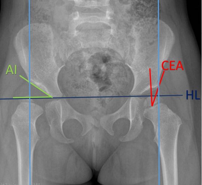
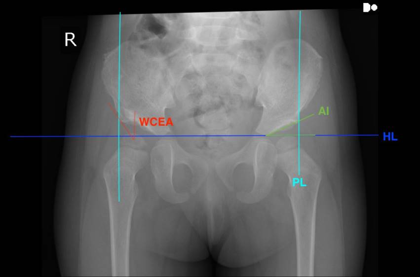
ÖZ Amaç: Gelişimsel Kalça Displazisi (GKD) olan hastalarda erken tanı ve erken tedavi önemlidir. Geç başvuran veya ihmal edilen hastalarda çeşitli osteotomi prosedürleri gerekebilir. Literatürde GKD cerrahisinde Pembersal osteotomi ile ilgili sınırlı sayıda makale bulunmaktadır. Çalışmamızın amacı GKD'li 18 aylıktan büyük çocuklarda izole Pembersal osteotominin sonuçlarını değerlendirmektir. Gereç ve Yöntemler: Bu retrospektif çalışmada 2014-2021 yılları arasında kliniğimizde GKD nedeniyle Pembersal osteotomi uygulanan hastaların sonuçları incelendi. Çalışmaya açık redüksiyon ile birlikte izole periasetabular Pembersal osteotomi uygulanan, en az 2 yıllık takibi olan ve yaşları 19 ile 36 ay arasında değişen 9 çocuk (7 kız, 2 erkek) dahil edildi. Ameliyat öncesi, ameliyat sonrası erken dönem, ameliyat sonrası 6. hafta ve son kontrol radyografileri değerlendirildi ve Wiberg'in orta kenar açısı (CEA), asetabular indeks (AI) ve Tönnis dereceleri not edildi. Bulgular: Hastaların ortalama yaşı 28.75 ay ve takip süresi 50.33 aydı. CEA ve AI ameliyattan sonra erken dönemde düzelme görüldü ve bu düzelme daha sonraki dönemlerde de devam ettiği görüldü. Tönnis evrelemesi tüm hastalarda ameliyattan sonra iyileşme gösterdi ve bu iyileşme de ameliyat sonrası erken dönemde ve 6. hafta kontrol grafilerde de devam etti. Ancak, son kontrol radyografilerinde 3 kalçada CEA<20º, AI>20º ve Tönnis evresi ≥2 idi ve bu üç kalça radyolojik kötü sonuç olarak değerlendirildi. Sonuç: Bu çalışma, izole Pembersal osteotominin gelişimsel kalça displazisinin tedavisinde asetabular indeks ve diğer radyolojik parametrelerde iyileşme ile birlikte etkili ve güvenli bir seçenek olduğunu göstermiştir. Anahtar Kelimeler: Gelişimsel kalça displazisi, periasetabular osteotomi, Pembersal osteotomi, merkez kenar açısı, asetabular indeks
Cite this Article As :
Baymurat AC, Abdulaliyev F, Ahmadov A, Osmanli E, Ok N, Gul O, Turanli S. Early and Mid-Term Radiological Results of Isolated Pembersal Procedure in Cases of Developmental Dislocation of The Hip. Selcuk Med J 2024;40(1): 16-21
1. Bakarman K, Alsiddiky AM, Zamzam M, et al. Developmental Dysplasia of the Hip (DDH): Etiology, Diagnosis, and Management. Cureus. 2023; 15:43207.
2. Ponseti IV. Growth and development of the acetabulum in the normal child. Anatomical, histological, and roentgenographic studies. J Bone Joint Surg Am. 1978; 60:575-85
3. Sungur I, Cetinus ME. Treatment of developmental dysplasia of the hip in children older than 18 months. TOTBID Journal 2014; 13:412-425.
4. Şenar M, Westhoff B, Zilkens C, et al. Indikation und Ergebnisse hüftnaher Osteotomien bei Dysplasie [Indications and results of corrective pelvic osteotomies in developmental dysplasia of the hip]. Orthopade. 2008; 37:556-70, 572-4, 576. German.
5. Agarwal A, Rastogi P. Clinicoradiological outcomes following pembersal acetabular osteotomy for developmental dysplasia of hip in young children: A series of 16 cases followed minimum 2 years. J Clin Orthop Trauma. 2021; 23:101669.
6. Bursali A, Tonbul M. How are outcomes affected by combining the Pemberton and Salter osteotomies? Clin Orthop Relat Res. 2008; 466:837-46.
7. Perlik PC, Westin GW, Marafioti RL. A combination pelvic osteotomy for acetabular dysplasia in children. J Bone Joint Surg Am. 1985; 67:842-50.
8. Li Y, Guo Y, Li M, et al. Multi-center Pediatric Orthopedic Study Group of China. Acetabular index is the best predictor of late residual acetabular dysplasia after closed reduction in developmental dysplasia of the hip. Int Orthop. 2018; 42:631-640.
9. Weinstein SL, Mubarak SJ, Wenger DR. Developmental hip dysplasia and dislocation: Part I. Instr Course Lect. 2004; 53:523-30.
10. Wenger DR, Bomar JD. Human hip dysplasia: evolution of current treatment concepts. J Orthop Sci. 2003; 8:264-71.
11. Akman B, Ozkan K, Cift H, et al. Treatment of Tönnis type II hip dysplasia with or without open reduction in children older than 18 months: a preliminary report. J Child Orthop. 2009; 3:307-11.
12. Albayrak M, Ugur F. Evaluating the effectiveness of interventions targeting only the acetabular side in the surgical treatment of Legg-Calvé-Perthes disease. Eur Rev Med Pharmacol Sci. 2023; 27:6151-6161.
13. Bortulev PI, Baskaeva TV, Vissarionov SV, et al. The choice of pelvic osteotomy technique in young children with hip dysplasia. Pediatric Traumatology, Orthopaedics and Reconstructive Surgery 2023; 11:5-16.
14. Sankar WN, Young CR, Lin AG, et al. Risk factors for failure after open reduction for DDH: a matched cohort analysis. J Pediatr Orthop. 2011; 31:232-9.



 Keywords