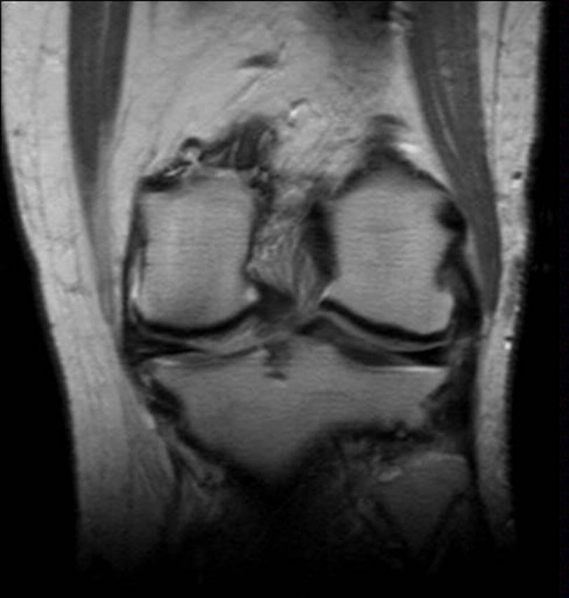
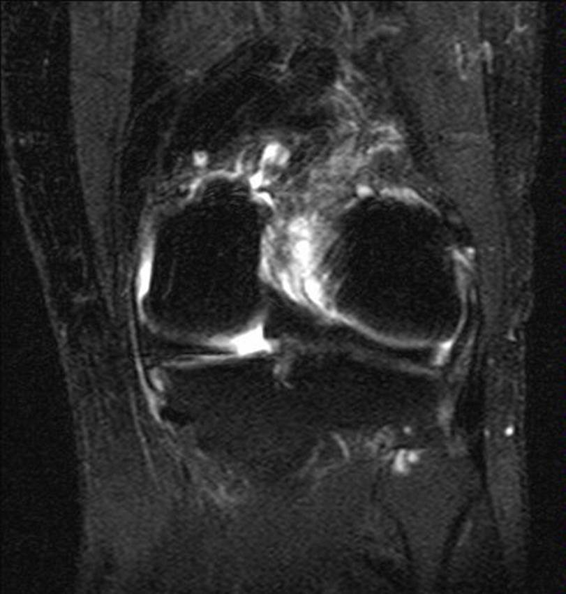
Manyetik Rezonans Görüntülemede Medial Menisküs Ekstrüzyonu: Yüksek Riskli Yırtıklar ve Eşlik Eden İntra-artiküler Patolojiler için Kritik Bir Gösterge
Amaç: Medial menisküs (MM) yırtık morfolojisi ile menisküs ekstrüzyonu arasındaki ilişkiyi ve ekstrüzyonun yüksek riskli yırtıkların belirlenmesinde bir gösterge olarak
kullanılıp kullanılamayacağını diz MRG bulguları üzerinden değerlendirmek.
Gereç ve Yöntemler: Bu retrospektif çalışmaya 160 hastaya ait 183 diz değerlendirmesi dahil edilmiştir. MM patolojileri dejenerasyon, horizontal-oblik, kök bölgesinde
radyal yırtık ve kompleks yırtık olarak sınıflandırılmıştır. Lateral menisküs patolojileri, kondromalazi, efüzyon ve ön çapraz bağ (ÖÇB) durumu kaydedilmiştir. Menisküs
ekstrüzyonu koronal proton-ağırlıklı kesitlerde ölçülmüştür. Gruplar arası farklar Ki-kare ve Kruskal-Wallis testleri ile değerlendirilmiş; yüksek riskli yırtıkları öngörmek
için ROC analizi yapılmıştır. Multivaryant lojistik regresyon modeli ≥3 mm ekstrüzyonun bağımsız belirleyicilerini araştırmıştır.
Bulgular: Radyal kök yırtığı ve kompleks yırtıklar diğer patolojilere göre belirgin derecede daha yüksek ekstrüzyon göstermiştir. Kondromalazi ve efüzyon, ≥3 mm
ekstrüzyonu olan dizlerde anlamlı olarak daha sık görülmüştür. ÖÇB rüptürü, hem yüksek ekstrüzyon hem de kompleks MM yırtıkları ile ilişkili bulunmuştur. ROC
analizinde yüksek riskli yırtıkları ayırt etmek için 3,27 mm (klinik olarak 3 mm) eşik değeri belirlenmiştir (duyarlılık %84,4; özgüllük %70,2; AUC 0,82). Regresyon
analizinde düşük-risk ve yüksek-risk yırtık grupları ile kondromalazi, patolojik ekstrüzyonun bağımsız belirleyicileri olarak bulunmuştur.
Sonuç: MM ekstrüzyonu özellikle 3 mm ve üzerindeki değerlerde, radyal kök ve kompleks yırtıkların güçlü bir radyolojik göstergesidir ve eşlik eden dejeneratif
değişiklikleri yansıtır. Ekstrüzyonun nicel değerlendirilmesi, cerrahi açıdan önemli yırtıkların erken tanısına katkı sağlayabilir.
Cite this Article As :
Gunbey Karabekmez L, Kerimoglu U, Gumus M, Akcam S, Tuncay I. Medial Meniscus Extrusion on MRI: A Critical Indicator for High-Risk Tears and
Associated Intra-Articular Pathologies. Selcuk Med J 2026;42(3): 224-233
1. Fox MG. MR Imaging of the Meniscus: Review, Current Trends, and Clinical Implications [Review of MR Imaging of the Meniscus: Review, Current Trends, and Clinical Implications]. Magnetic Resonance Imaging Clinics of North America. 2007;15(1):103. doi:10.1016/j.mric.2007.02.004.
2. Shapiro LM, Staroswiecki E, Gold GE. Magnetic Resonance Imaging of the Knee: Optimizing 3 Tesla Imaging [Review of Magnetic Resonance Imaging of the Knee: Optimizing 3 Tesla Imaging]. Seminars in Roentgenology. 2010;45(4):238. doi:10.1053/j.ro.2009.12.007.
3. Vaidya S, Aneesh MK, Mahajan SM, et al. Radiological Assessment of Meniscal Injuries of the Knee on Magnetic Resonance Imaging. International Journal of Current Research and Review. 2020;12(15):98. doi:10.31782/ijcrr.2020.121511.
4. Cao J, Chen B. Function, Injury, and Treatment for Meniscus. Highlights in Science Engineering and Technology. 2022;8:263. doi:10.54097/hset.v8i.1145.
5. McDermott I, Masouros SD, Amis AA. Biomechanics of the menisci of the knee. Current Orthopaedics. 2008;22(3):193. doi:10.1016/j.cuor.2008.04.005.
6. Luvsannyam E, Jain MS, Leitao AR, et al. Meniscus Tear: Pathology, Incidence, and Management [Review of Meniscus Tear: Pathology, Incidence, and Management]. Cureus. 2022. doi:10.7759/cureus.25121.
7. Bo K, Liu X, Zhang Y, et al. Associations between the meniscal lesion parameters and knee joint structures and symptoms in patients with symptomatic knee osteoarthritis. Arthritis Research & Therapy. 2025;27(1). doi:10.1186/s13075-025-03583-y.
8. Nie S, Li H, Jing-ping G, et al. Association between anatomical risk factors and medial meniscus posterior root tears: a retrospective study. BMC Musculoskeletal Disorders. 2025;26(1). doi:10.1186/s12891-025-08676-y.
9. Olivotto E, Trisolino G, Belluzzi E, et al. Macroscopic Synovial Inflammation Correlates with Symptoms and Cartilage Lesions in Patients Undergoing Arthroscopic Partial Meniscectomy: A Clinical Study. Journal of Clinical Medicine. 2022;11(15):4330. doi:10.3390/jcm11154330.
10. Kim DH, Lee GC, Kim HH, et al. Correlation between meniscal extrusion and symptom duration, alignment, and arthritic changes in medial meniscus posterior root tear: research article. Knee Surgery and Related Research. 2019;32(1). doi:10.1186/s43019-019-0019-x.
11. Crema MD, Roemer FW, Felson DT, et al. Factors Associated with Meniscal Extrusion in Knees with or at Risk for Osteoarthritis: The Multicenter Osteoarthritis Study. Radiology. 2012;264(2):494. doi:10.1148/radiol.12110986.
12. Rennie WJ, Finlay D. Meniscal Extrusion in Young Athletes: Associated Knee Joint Abnormalities. American Journal of Roentgenology. 2006;186(3):791. doi:10.2214/ajr.04.1181.
13. Papalia GF, Zà P, Saccone L, et al. Meniscal extrusion: risk factors and diagnostic tools to predict early osteoarthritis. Orthopedic Reviews. 2023;15. doi:10.52965/001c.74881.
14. Hada S, Ishijima M, Kaneko H, et al. Association of medial meniscal extrusion with medial tibial osteophyte distance detected by T2 mapping MRI in patients with early-stage knee osteoarthritis. Arthritis Research & Therapy. 2017;19(1). doi:10.1186/s13075-017-1411-0.
15. Kan A, Oshida M, Oshida S, et al. Anatomical significance of a posterior horn of medial meniscus: the relationship between its radial tear and cartilage degradation of joint surface. BMC Sports Science Medicine and Rehabilitation. 2010;2(1). doi:10.1186/1758-2555-2-1.
16. Koo JH, Choi SH, Lee SA, et al. Comparison of Medial and Lateral Meniscus Root Tears. PLoS ONE. 2015;10(10). doi:10.1371/journal.pone.0141021.
17. Bloecker K, Wirth W, Guermazi A, et al. Relationship Between Medial Meniscal Extrusion and Cartilage Loss in Specific Femorotibial Subregions: Data From the Osteoarthritis Initiative. Arthritis Care & Research. 2015;67(11):1545. doi:10.1002/acr.22615.
18. Hada S, Kaneko H, Liu L, et al. Medial meniscus extrusion is directly correlated with medial tibial osteophyte in patients received reconstruction surgery for anterior cruciate ligament injury: A longitudinal study. Osteoarthritis and Cartilage Open. 2022;4(4):100320. doi:10.1016/j.ocarto.2022.100320.
19. Ma X, Liu Q, Xu D, et al. Biomechanical impact of progressive meniscal extrusion on the knee joint: a finite element analysis. Journal of Orthopaedic Surgery and Research. 2024;19(1). doi:10.1186/s13018-024-05249-y.
20. Makiev KG, Vasios IS, Georgoulas P, et al. Clinical significance and management of meniscal extrusion in different knee pathologies: a comprehensive review of the literature and treatment algorithm [Review of Clinical significance and management of meniscal extrusion in different knee pathologies: a comprehensive review of the literature and treatment algorithm]. Knee Surgery and Related Research. 2022;34(1). doi:10.1186/s43019-022-00163-1.
21. Li L, Yang X, Yang L, et al. Biomechanical analysis of the effect of medial meniscus degenerative and traumatic lesions on the knee joint. PubMed. 2019;11(2):542. PMID: 30899361; PMCID: PMC6413253. https://pubmed.ncbi.nlm.nih.gov/30899361.
22. Costa C, Morrison WB, Carrino JA. Medial Meniscus Extrusion on Knee MRI: Is Extent Associated with Severity of Degeneration or Type of Tear? American Journal of Roentgenology. 2004;183(1):17. doi:10.2214/ajr.183.1.1830017.
23. Allen DM, Li L, Crema MD, et al. The relationship between meniscal tears and meniscal position. Therapeutic Advances in Musculoskeletal Disease. 2010;2(6):315. doi:10.1177/1759720x10383198.
24. Lee D, Lee B, Kim J, et al. Predictors of degenerative medial meniscus extrusion: radial component and knee osteoarthritis. Knee Surgery Sports Traumatology Arthroscopy. 2010;19(2):222. doi:10.1007/s00167-010-1274-2.
25. Muzaffar N, Kirmani O, Ahsan MS, et al. Meniscal Extrusion in the Knee: Should only 3 mm Extrusion be Considered Significant? An Assessment by MRI and Arthroscopy. Malaysian Orthopaedic Journal. 2015;9(2):17. doi:10.5704/moj.1507.013.
26. Magee T. Usefulness of Unenhanced MRI and MR Arthrography of the Shoulder in Detection of Unstable Labral Tears. American Journal of Roentgenology. 2015;205(5):1056. doi:10.2214/ajr.14.14262.
27. Gajjar SM, Solanki K, Shanmugasundaram S, et al. Meniscal Extrusion: A Narrative Review [Review of Meniscal Extrusion: A Narrative Review]. Orthopaedic Journal of Sports Medicine. 2021;9(11). doi:10.1177/23259671211043797.
28. Choi CJ, Choi YJ, Lee JJ, et al. Magnetic Resonance Imaging Evidence of Meniscal Extrusion in Medial Meniscus Posterior Root Tear. Arthroscopy The Journal of Arthroscopic and Related Surgery. 2010;26(12):1602. doi:10.1016/j.arthro.2010.05.004.
29. Furumatsu T, Kintaka K, Higashihara N, et al. Meniscus extrusion is a predisposing factor for determining arthroscopic treatments in partial medial meniscus posterior root tears. Knee Surgery and Related Research. 2023;35(1). doi:10.1186/s43019-023-00182-6.
30. Daszkiewicz K, Łuczkiewicz P. Biomechanics of the medial meniscus in the osteoarthritic knee joint. PeerJ. 2021;9. doi:10.7717/peerj.12509.
31. Lee SY, Jee WH, Kim JM. Radial Tear of the Medial Meniscal Root: Reliability and Accuracy of MRI for Diagnosis. American Journal of Roentgenology. 2008;191(1):81. doi:10.2214/ajr.07.2945.
32. Krych AJ, LaPrade MD, Hevesi M, et al. Investigating the Chronology of Meniscus Root Tears: Do Medial Meniscus Posterior Root Tears Cause Extrusion or the Other Way Around? Orthopaedic Journal of Sports Medicine. 2020;8(11). doi:10.1177/2325967120961368.
33. Xu ZH, Li Y, Rao J, et al. Biomechanical assessment of disease outcome in surgical interventions for medial meniscal posterior root tears: a finite element analysis. BMC Musculoskeletal Disorders. 2022;23(1). doi:10.1186/s12891-022-06069-z.
34. Moon H, Choi C, Jung M, et al. Medial Meniscus Posterior Root Tear: How Far Have We Come and What Remains? [Review of Medial Meniscus Posterior Root Tear: How Far Have We Come and What Remains?]. Medicina. 2023;59(7):1181. doi:10.3390/medicina59071181.
35. LaPrade CM, Foad A, Smith SD, et al. Biomechanical consequences of a nonanatomic posterior medial meniscal root repair. Am J Sports Med. 2015;43(4):912-920. doi:10.1177/0363546514564150.
36. Srikanth VK, Fryer JL, Zhai G, et al. A meta-analysis of sex differences in prevalence, incidence and severity of osteoarthritis. Osteoarthritis Cartilage. 2005;13(9):769-781. doi:10.1016/j.joca.2005.04.014.
37. Dunn WR, Spindler KP. Predictors of activity level 2 years after anterior cruciate ligament reconstruction (ACLR): a multicenter Orthopaedic Outcomes Network (MOON) ACLR cohort study. Am J Sports Med. 2010;38(10):2040-2050. doi:10.1177/0363546510370283.
 Research Paper
 View as PDF
 Keywords
Menisküs yırtığı, menisküs ekstrüzyonu, MRG, Ön Çapraz Bağ yırtığı, kondromalazi
Keywords
Menisküs yırtığı, menisküs ekstrüzyonu, MRG, Ön Çapraz Bağ yırtığı, kondromalazi